TAPP INGUINAL HERNIA REPAIR
TAPP Inguinal Hernia Repair
Patient Selection for TAPP Inguinal Hernia Repair
Difficult cases:
- Indirect hernial sacs are closely applied to the cord structures and are more often complete, making dissection difficult.
- Left-sided hernias are more difficult to dissect than the right-sided ones for a beginner.
- Recurrent hernias and irreducible hernias.
- Obese patients.
Direct or small indirect primary hernias in lean and thin subjects are the best during learning curve.
Anesthesia
General anesthesia
Skin Preparation
No studies are there assessing preparation for hernia surgery, but as with other surgical procedures, there is no difference in surgical site infections (SSI) in preparation and no preparation group. In
our centre, we prepare skin of all male patients before surgery.
Catheterization of the patient
The patient is asked to pass urine just before shifting him to the operation theater. Predisposing factors for an injury are a full bladder or a previous exposure of the retropubic space, particularly after prostate interventions, irradiation, or in case of huge or recurrent hernias (16). If the patient has any of these factors, Foley’s indwelling catheter is placed prior to surgery. It may be removed the next morning.
Antibiotic prophylaxis
Hernia repair is considered a clean surgery (infection rate <2%). With age >75 years, obesity, urinary catheter, recurrent hernia, diabetes, immunosuppressant/ corticosteroid usage, and malignancy, infection rate rises to 14%(17). In endoscopic repair, antibiotic prophylaxis does not significantly reduce the number of wound infections- level 2B evidence. In the presence of risk factors for wound infection based on patient factors (recurrence, advanced age, immunosuppressive conditions) or surgical factors (expected long operating times, use of drains), the use of antibiotic prophylaxis should be considered – Grade C recommendation. But in our center, preoperative prophylactic antibiotics are administered (ceftriaxone & sulbactam at induction and tobramycin two hours prior) for all.
After induction of anesthesia, irreducible hernial contents, if any, are reduced if possible before painting & draping is commenced.
The patient lies supine with both arms tucked by the side, to make room for the surgeon and his assistant to stand at shoulder level. The monitor is positioned at the foot end of the patient. The operating surgeon stands on the side opposite to hernia. The assistant, who holds the camera, stands on the side of hernia or behind the surgeon. The scrub nurse positions herself to the left of the patient, to the left of the surgeon.
Pneumoperitoneum and placement of ports
Open umbilical or Palmer’s point entry: Direct open entry with blunt trocar is as safe as Veress needle entry, level 1B evidence.
A vertical umbilical incision is made. The abdominal wall is lifted up and stabilized with one hand and the 10-mm blunt trocar is directed towards the hollow of the pelvis. A 0 degree telescope attached to the camera is introduced and the entry point inspected. The head end of the table is kept 20-300 low to facilitate movement of the bowel away from the operative field. The groin area is then visualized. Two 5-mm ports are placed as working ports for the right and left hand of the surgeon, one on each side, at the level of umbilicus in the midclavicular line. These ports should be placed under vision to prevent injury to the inferior epigastric vessels and underlying bowel. The telescope is now changed to 30 degrees for better view.
The hernia defect is inspected and the type of hernia (direct or indirect) is confirmed by the position of defect in relation to the inferior epigastric vessels and cord structures. The spermatic vessels will be seen laterally and the vas deferens medially and they meet at the internal ring (round ligament instead of vas deferens in female). This forms an inverted V. The inferior epigastric vessels (IEV) can be seen coursing upwards from this point. A direct hernia is medial to the IEV. An indirect hernia is lateral to the IEV.

Fig.3.3- TAPP Inguinal Hernia Repair – Anatomical landmarks, right side, indirect defect

Fig. 3.4 – TAPP Inguinal Hernia Repair – Direct hernia, left side.
All the anatomical landmarks normally seen before peritoneal reflection are identified. These include the median umbilical ligament in the midline, the medial umbilical ligaments on each side and the lateral umbilical ligaments. Contents of the hernial sac, if any, are reduced with the help of atraumatic bowel forceps. In case of irreducible hernias, the bowel contents need to be handled with care. In case of omentum, it may be reduced or excised and removed via 2-cm scrotal incision.
Operative Steps
Step 1– Incising the peritoneum
The peritoneal incision is begun at a point that is midway between the groin crease and the umbilicus, generally about 4-5 cm above all the defects. It may be worth marking the line of proposed peritoneal cut using an energy source so as to avoid coming closer to the defect later. Incision on the peritoneum is made from medial to lateral i.e. from right side to the left on the left side and from left side to right on the right side. Scissors, monopolar hook, or harmonic scalpel can be used for the dissection. It extends from the medial umbilical ligament to the paracolic gutter. Extending it medially beyond the medial umbilical ligament may increase the chances of injury to the urinary bladder, particularly if the urinary bladder is not empty especially if the peritoneal cut is not placed towards umbilicus.
Step 2 – Raising the peritoneal flap
The correct plane of dissection of the peritoneal flap from the transversus muscle is anterior to the preperitoneal fascia through the loose areolar tissue, i.e. in the space of Bogros. The flap is raised by both blunt and sharp dissection. Generally, the plane is avascular, but any small vessel is carefully cauterized before division.
Care should be taken to avoid injury to the inferior epigastric vessels (IEV) while raising the peritoneum medial to the internal ring. These vessels should always be left attached to the muscle and should never be included in the flap. Otherwise, they may come in the way of dissection and may get injured. The plane of dissection is easier on the medial side and blunt dissection is sufficient since the areolar tissue is loose and the peritoneum is not adhered to the rectus muscle. On the medial side, continued caudal dissection will identify the shiny Cooper’s ligament and the pubic bone. Laterally, the peritoneum is slightly adhered to the transversus muscle, and sharp dissection may be required. Take care not to get into the plane of muscles.
In case of local omental or intestinal adhesions to the peritoneum of the groin, it is not recommended to perform adhesiolysis in general, unless it obstructs the view. In sliding hernias or even irreducible hernias, neither adhesiolysis nor reduction is mandatory, but the straightforward preperitoneal dissection should be performed. This facilitates the mobilization of hernia content within the sac and helps to avoid intestinal injury.
Step 3 – Dissection of medial peritoneum and direct sac
Dissection is continued medially to the pubic symphysis to visualize the cave of Retzius. The medial dissection should go across the midline to the opposite side, up to opposite IEV; so that the mesh can be placed with a good overlap over the defect. A direct defect is encountered medially above the iliopubic tract. The direct sac can be easily pulled inside along with essentially thinned out fascia transversalis identified by its glistening appearance and belongs to the parietal wall, i.e., pseudosac. With sharp and blunt dissection, sac is separated from pseudosac (a push pull technique). In case of large direct hernias, after reducing the sac, the dome of the pseudosac can be fixed to the rectus muscle over the pubic bone with a few tacks to prevent postoperative seroma formation and also helps to have a base for the mesh, Level 2B evidence (18).
Step 4 – Lateral dissection
After the medial dissection, the flap is raised lateral to the internal ring till the anterior superior iliac spine is reached and it is carried posteriorly over the psoas muscle. Care is taken during this dissection to avoid injury to the nerves overlying the psoas muscle namely lateral cutaneous nerve of the thigh laterally and the femoral branch of the genitofemoral nerve medially.
Retroperitoneal area lateral to the cord structures on the left side. The lateral cutaneous nerve of the thigh (1) and the femoral branch of the genitofemoral nerve (2) can be seen coursing on the psoas muscle (3). Both the nerves enter the thigh below the iliopubic tract (4).
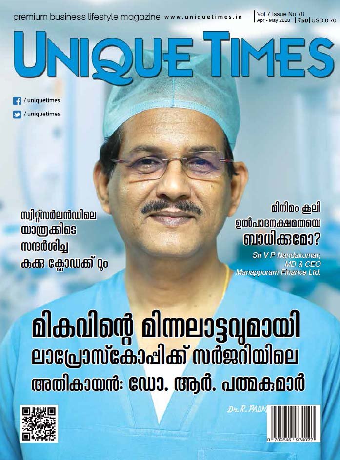
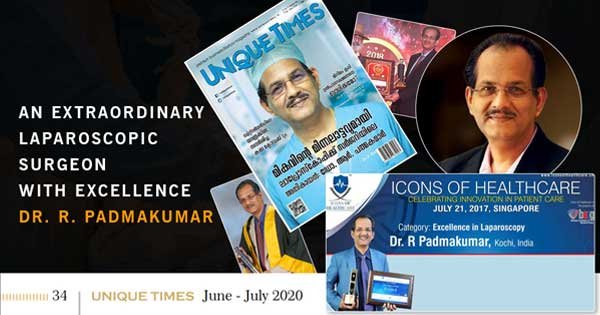
About Dr. R. Padmakumar
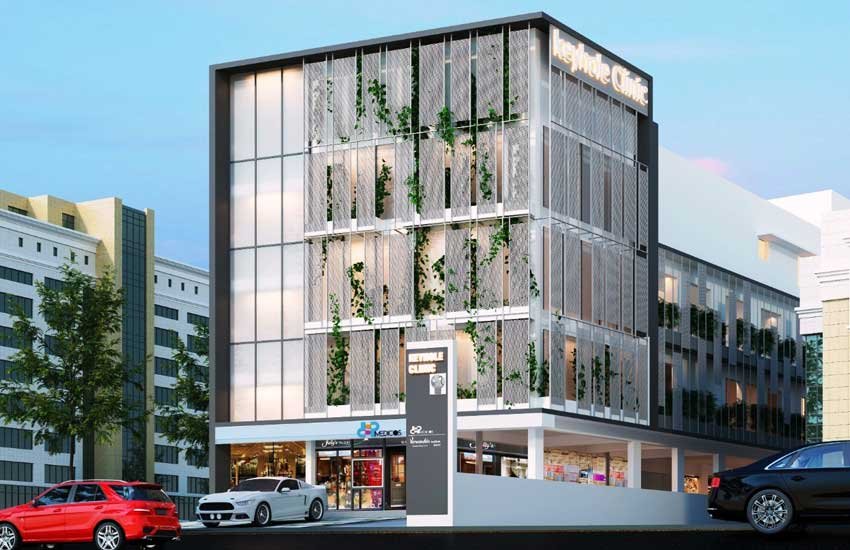
Dr. R. Padmakumar is one of the Best Hernia Surgeon in India. He has been changing lives through Keyhole Surgery. He has got more than 30 years of hands-on experience in laparoscopic Surgery in major hospitals across India and has completed more than 7000 cases of Laparoscopic Hernia Surgeries. Dr. Padmakumar has trained more than 300 surgeons from all over the world the art of laparoscopic surgery especially Laparoscopic Hernia Surgery. Dr. Padmakumar is also hailed as one of the Best Bariatric Surgeon in India & UAE. He is renowned for new and improved treatment techniques and the first in the World to perform Scarless Bariatric Surgery with Tummy Tuck / Abdominoplasty. He is also one of the very few thyroid surgeons in India doing Endoscopic Thyroidectomy (scarless thyroid surgery).


-
Anal Fissure Treatment
-
Gynaecomastia Treatment
- Piles / Hemorrhoids Treatment
- Varicose Veins Treatment
- Laser Treatment for Varicose Veins
- Diabetic Foot Treatment


